Evolution and diversity of biomineralized columnar architecture in early Cambrian phosphatic-shelled brachiopods
- State Key Laboratory of Palaeobiology and Stratigraphy, Nanjing Institute of Geology and Palaeontology, Chinese Academy of Sciences, China
- School of Natural Sciences, Macquarie University, Australia
- State Key Laboratory of Continental Dynamics, Shaanxi Key Laboratory of Early Life & Environments, Department of Geology, Northwest University, China
- Institute of Earth Sciences, Palaeobiology, Uppsala University, Sweden
- Department of Palaeobiology, Swedish Museum of Natural History Stockholm, Sweden
Figures
Figure 1
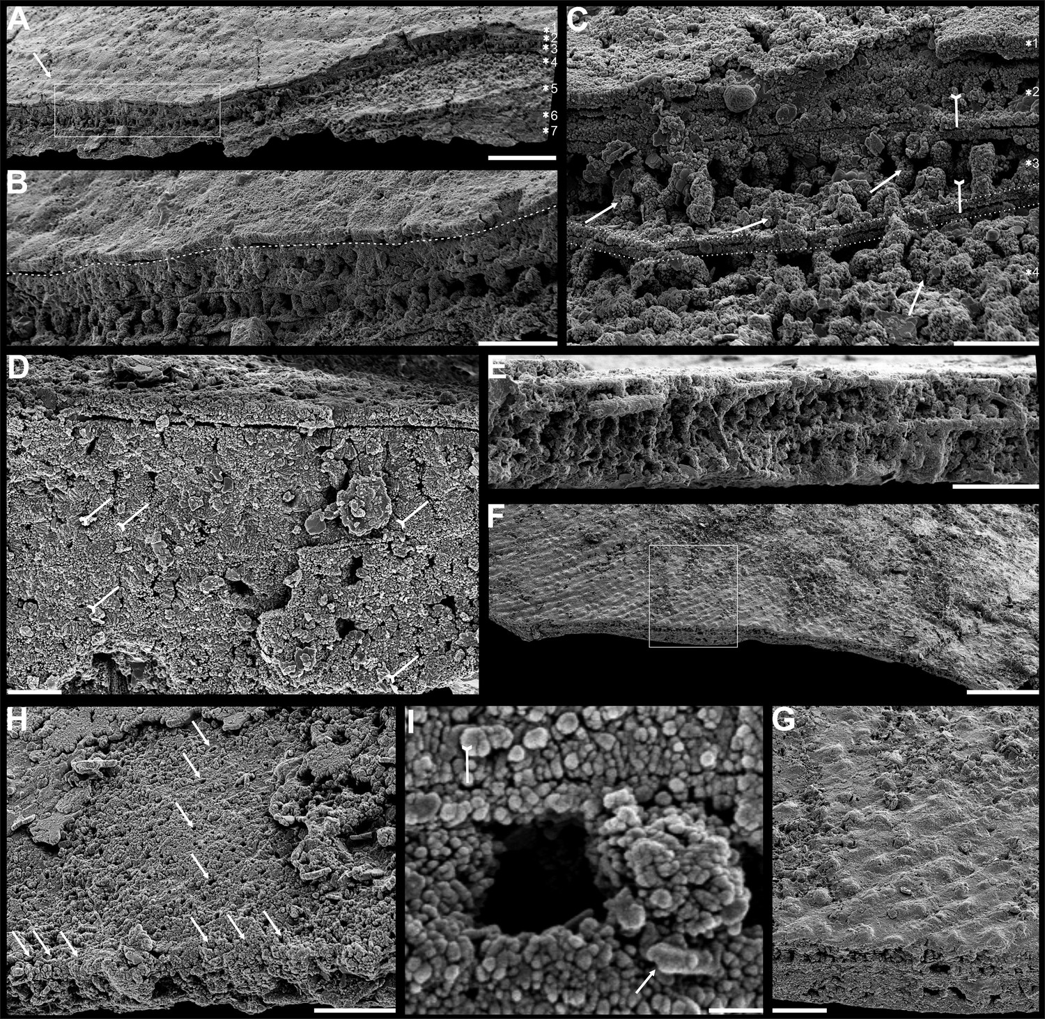
Shell architecture of Latusobolus xiaoyangbaensis gen. et sp. nov. from the Cambrian Series 2 Shuijingtuo Formation in southern Shaanxi, South China.
(A–C) ELI-XYB S5-1 BS01. (A) Cross-section of a ventral lateral margin, note post-metamorphic pustules by arrow, primary layer 1 and stacked sandwich columnar units 2–7, box indicates area in B. (B) Enlarged view of (A) showing the boundary between top primary and underlying secondary layers by dotted line. (C) Enlargement of shell layers 1–4 of (A) note canals of columns (arrows), gap (tailed arrows) between two stratiform lamellae by dotted lines. (D) Poorly phosphatised columns of ventral valve, note canals by tailed arrows, ELI-XYB S5-1 BR06. (E) Columns of dorsal valve, ELI-XYB S5-1 BS17. (F) Cross-section of a ventral lateral margin, showing post-metamorphic pustules, box indicates the area in (G), ELI-XYB S4-2 BO06. (G) Enlarged primary layer pustules and underlying secondary layer columns. (H) Dorsal valve, one unit of stacked columnar architecture with the exfoliation of top primary layer, noting column canals on the stratiform lamella surface by arrows, ELI-XYB S4-2 BO08. (I) Apatite spherules of granule aggregations of ventral columnar shell structure, note granule rods by arrow and thin gap left by the degradation of organic counterparts by tailed arrow, ELI-XYB S4-2 BO06. Scale bars: (A), 50 µm; (B), (E), (G), 20 µm; (C), (H), 10 µm; (D), 5 µm; (F), 100 µm; (I), 1 µm.
Figure 2
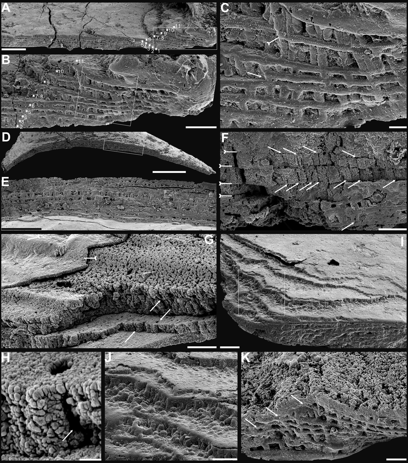
Shell architecture of ventral Eoobolus acutulus sp. nov. from the Cambrian Series 2 Shuijingtuo Formation in Three Gorges areas, South China.
(A–C) ELI-AJH S05 BT12. (A) Cross-section of a ventral lateral margin, note primary layer 1 and stacked sandwich columnar units 2–11, box indicates area in B. (B) Enlarged view of A. (C) Enlarged view of B, show thin gap left by the degradation of organic counterparts (tailed arrow) between two stratiform lamellae by dotted lines, the fusion point of two columnar units by arrow. (D–F) ELI-AJH 8-2-3 BT02. (D) Cross-section of shell margin, box indicates area in E. (E) Different preservation condition of columnar architecture. (F) Poorly phosphatised columns, note the opening of canals along the organic membrane by arrows, and space between two stratiform lamellae by tailed arrows. (G–H) ELI-AJH 8-2-3 BT03. (G) Note canals on the cross-section and surface of stratiform lamella by arrows, and partly exfoliated primary layer by tailed arrow. (H) Magnified columns in (G), composed of granule spherules with canal by arrow. (I) Cross-section of shell margin, box indicates area in (J), ELI-AJH 8-2-3 BT04. (J) Enlarged short columns. (K) Imbricated columnar architecture (arrows), ELI-AJH S05 BT12. Scale bars: (A), (E), 50 µm; (B), (I), 20 µm; (C), 5 µm; (D), 200 µm; (F), (G), (J), (K), 10 µm; (H), 1 µm.
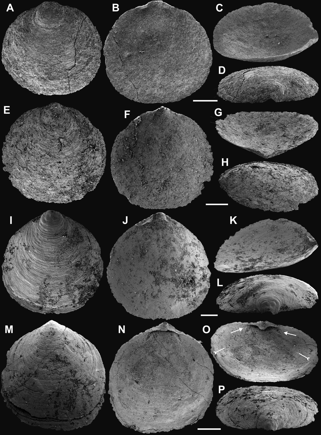
Figure 3
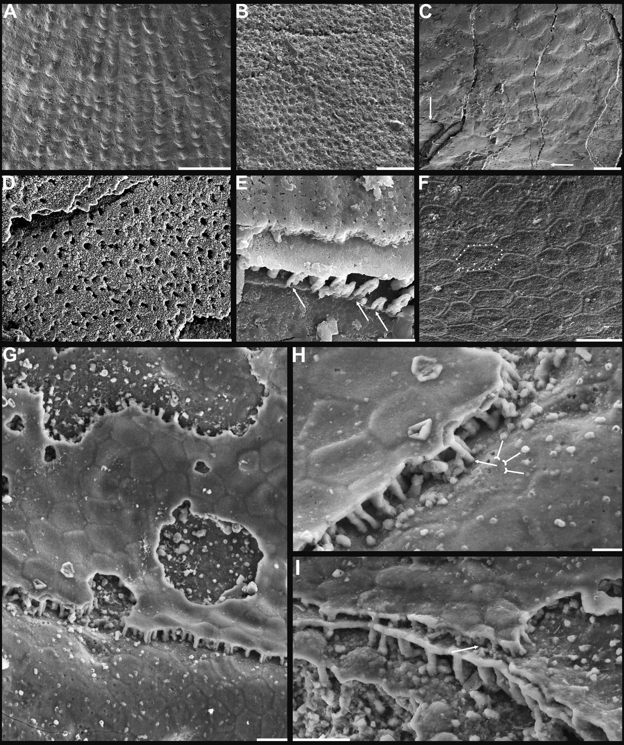
Shell ornamentation, ultrastructure, and epithelial cell moulds of Cambrian Series 2 brachiopods.
(A) Post-metamorphic pustules of Latusobolus xiaoyangbaensis gen. et sp. nov., ELI-XYB S4-3 AU11. (B–D) Eoobolus acutulus sp. nov. (B) Metamorphic hemispherical pits, ELI-AJH 8-2-2 Lin01. (C) Epithelial cell moulds, note column openings on layer surfaces beneath by arrows, ELI-AJH S05 N31. (D) Enlarged column openings on layer surface, ELI-WJP 7 CE05. (E–I) Eohadrotreta zhenbaensis. (E) Partly broken columns, note canals by arrows, ELI-AJH F36. (F) Polygonal epithelial cell moulds on valve floor, dotted line indicates margin of one epithelial cell, ELI-WJP 6 R79. (G–I) ELI-AJH Acro 053. (G) Epithelial cell moulds on dorsal median septum with columns between them. (H) Enlarged view of G, note rudiment of columns by tailed arrows and one column on epithelial cell margin by arrow. (I) Epithelial cell moulds on stratiform lamella surfaces of successive three stacked sandwich columnar units developed on cardinal muscle areas with columns between (marked by arrow). Scale bars: (A), (D), 50 µm; (B), (E), (F), (H), 10 µm; (C), (G), (I), 20 µm.
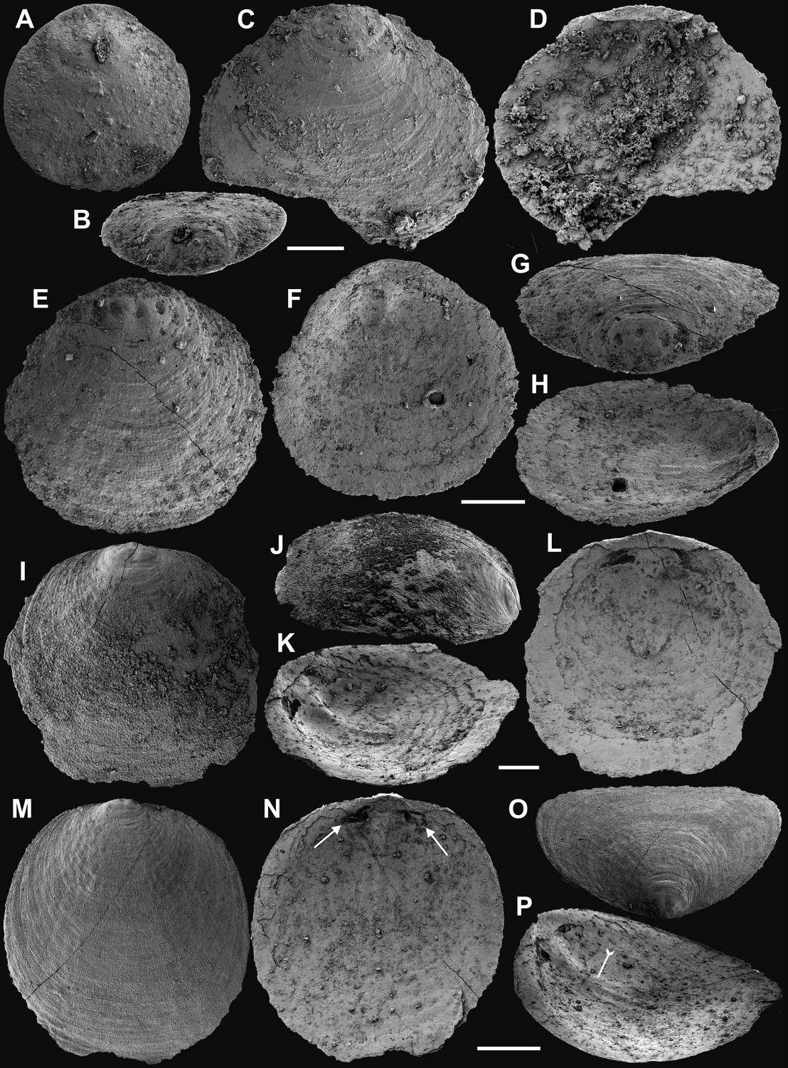
Figure 4
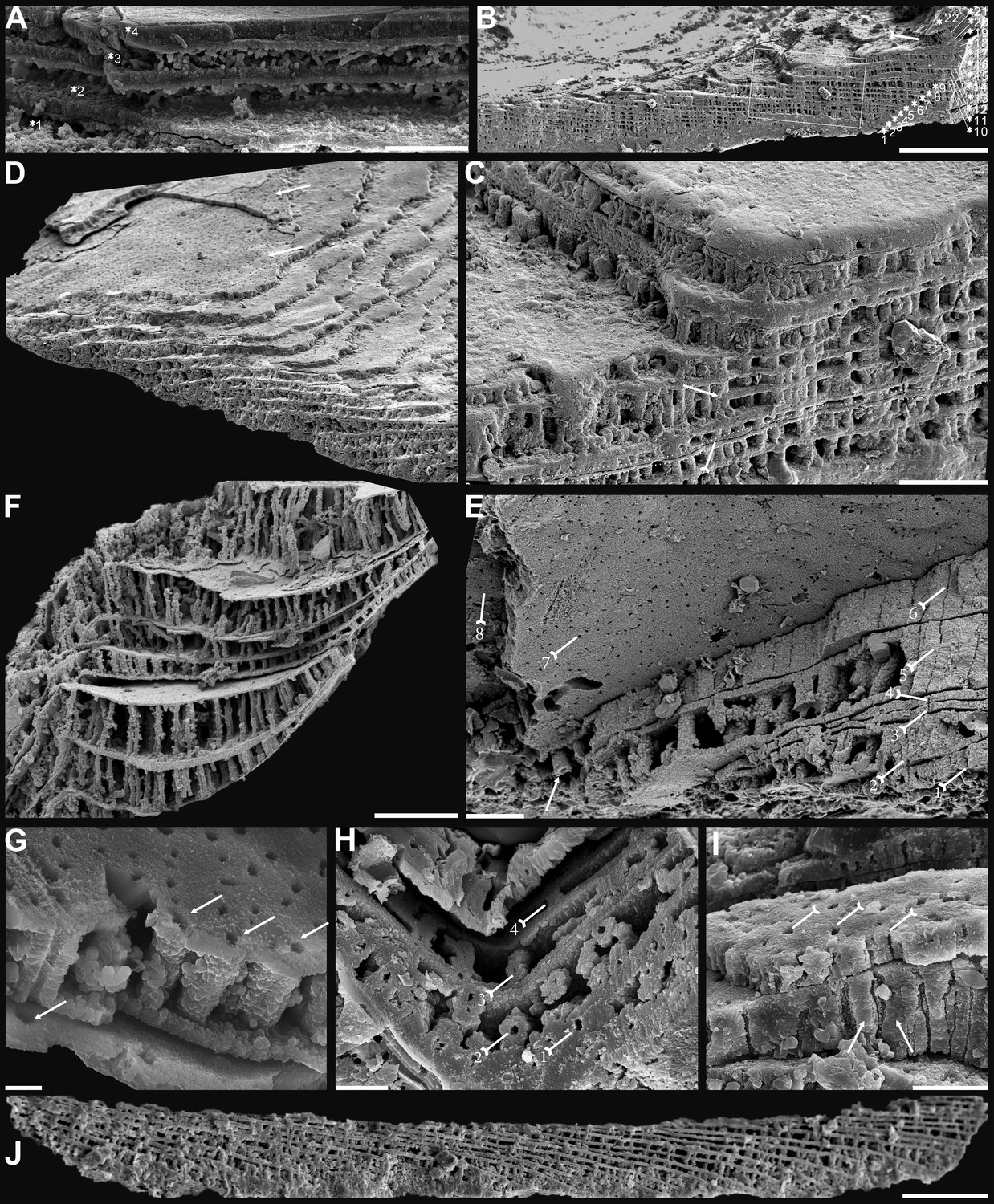
Biomineralized columnar architecture of Cambrian Series 2 brachiopods.
(A) Eoobolus incipiens, P00712-AJXM-267.5 DT-12. (B–D) Lingulellotreta ergalievi, ELI-AJH 8-2-3 CI11. (B) Cross-section of shell margin, box indicates area in C, note primary layer 1 and stacked sandwich columnar units 2–22, and raised pseudointerarea (tailed arrow). (C) Enlarged view of thin gap (tailed arrow) between two stratiform lamellae by dotted lines, the fusion of two stacked columnar units into one by arrow. (D) Imbricated growth pattern of stacked columnar units. (E) Palaeotreta zhujiahensis, note column openings (arrow) on eight successive columnar units by tailed arrows, ELI-AJH 8-2-1 AE09. (F–J) Eohadrotreta zhenbaensis. (F) Relatively taller columns (ca. 20 µm), ELI-AJH 8-2-1 acro16. (G) Apatite spherules of granule aggregations in one columnar unit, note column openings (arrows) on both stratiform lamella surfaces, ELI-AJH S05 E18. (H) Cross-section shows column openings on four successive units by tailed arrows, ELI-WJP 7 AB98. (I) Poorly phosphatised columns (arrows), note openings of canals on surface of stratiform lamella by tailed arrows, ELI-AJH S05 I76. (J) Stacked columnar units in an imbricated pattern, ELI-WJP 6 R47. Scale bars: (A), (E), (I), 10 µm; (B), 100 µm; (C), (F), 20 µm; (G), 2 µm; (H), 5 µm; (D), (J), 50 µm.
Figure 5
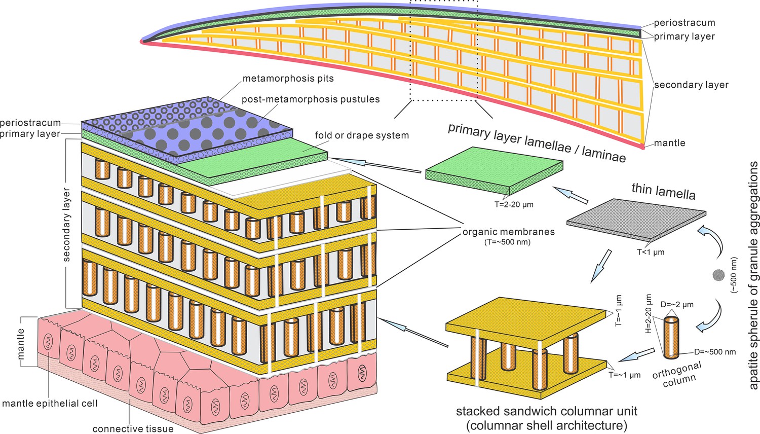
Biomineralization process of typical columnar architecture using the stacked sandwich model of phosphatic-shelled brachiopods.
Abbreviation: D=Diameter; H=height; T=Thickness. modified from Williams and Holmer, 1992 (Text-Fig. 7); Zhang et al., 2016b (Figure 6).
Figure 6
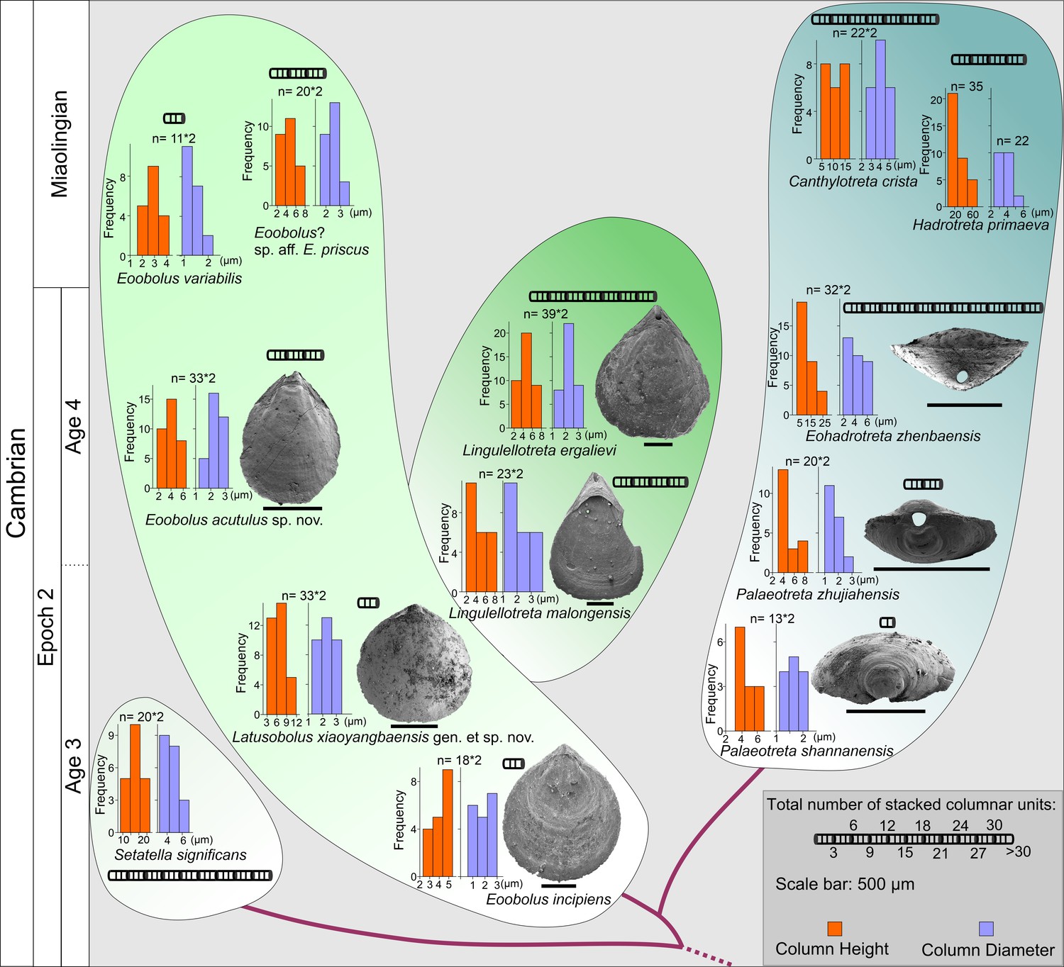
The evolution of stacked sandwich columnar architecture in early Eoobolidae taxa, Eoobolus incipiens, Latusobolus xiaoyangbaensis gen. et sp. nov., Eoobolus acutulus sp. nov., Eoobolus variabilis, Eoobolus? aff. priscus, Lingulellotretidae Lingulellotreta malongensis, Lingulellotreta ergelievi, Acrotretida Palaeotreta shannanensis, Palaeotreta zhujiahensis, Eoohadrotreta zhenbaensis, Hadrotreta primaeva, Canthylotreta crista, and stem group Setatella significans.
The height and diameter data of columns are based on data from literature (Skovsted and Holmer, 2003; Streng et al., 2007; Streng and Holmer, 2006; Ushatinskaya and Korovnikov, 2014; Zhang et al., 2016b; Zhang et al., 2020b; Zhang et al., 2020c).
-
Figure 6—source data 1
Raw data of the measurements of diameter and height of columns, thickness of different shell layers and number of columnar units demonstrated in Figure 6.
- https://cdn.elifesciences.org/articles/88855/elife-88855-fig6-data1-v1.xlsx
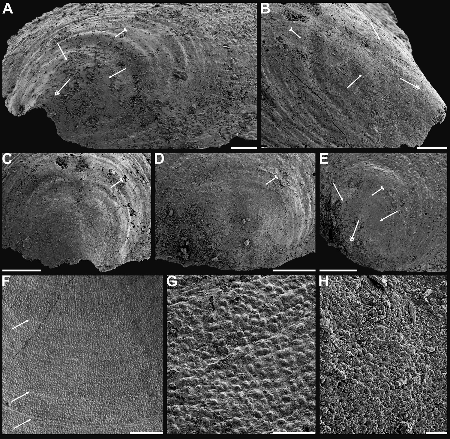
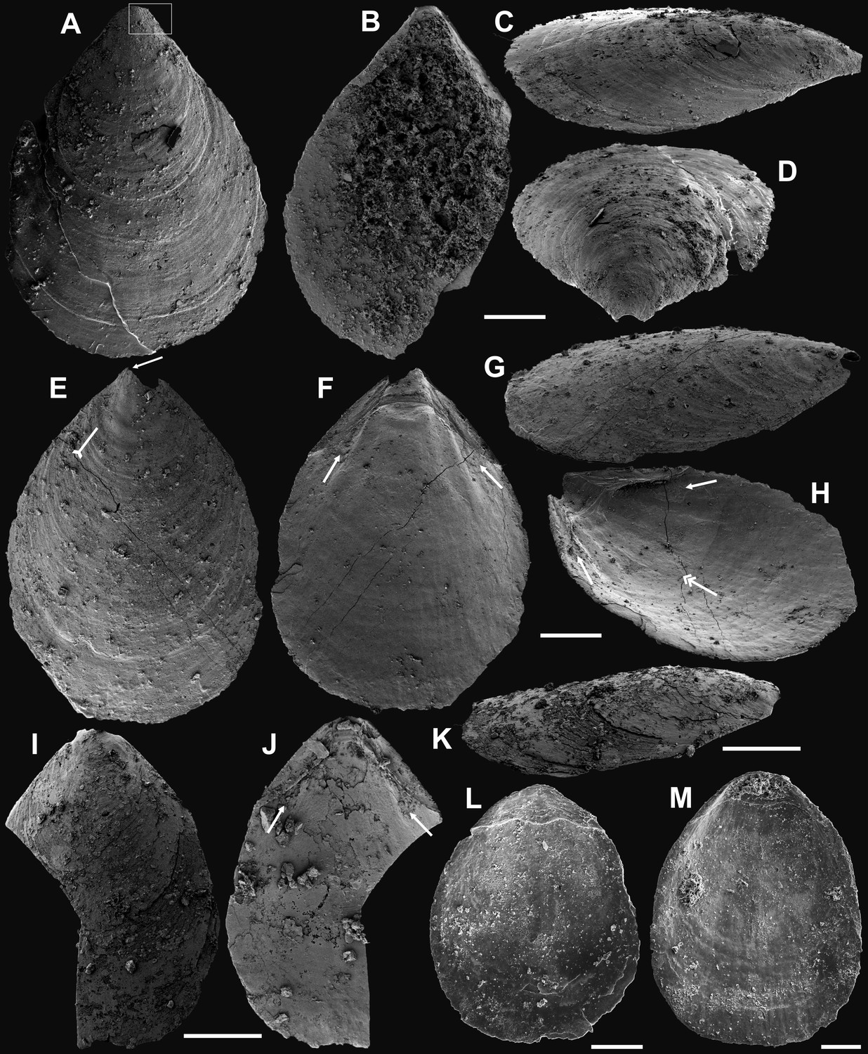
Appendix 1—figure 1
Ventral valves of Latusobolus xiaoyangbaensis gen. et sp. nov. from the Cambrian Series 2 Shuijingtuo Formation in southern Shaanxi, South China.
(A–D) Juvenile with weakly developed pseudointerarea, ELI-XYB S5-1 BR01. (E–H) Juvenile with weakly developed pseudointerarea, ELI-XYB S5-1 BR02. (I–J) Large valve with slightly elevated pseudointerarea, ELI-XYB S5-1 BR04. (M–P) Mature valve with developed posterolateral muscle scars (arrows) and rudiment of vascula lateralia (tailed arrows), ELI-XYB S5-1 BR09. Scale bars: (A–L), 200 µm; (M–P), 500 µm.
Appendix 1—figure 2
Dorsal valves of Latusobolus xiaoyangbaensis gen. et sp. nov. from the Cambrian Series 2 Shuijingtuo Formation in southern Shaanxi, South China.
(A–B) Small juvenile with weakly developed pustular ornamentation, ELI-XYB S5-1 BS15. (C–D) Juvenile, ELI-XYB S4-2 BO08. (E–H) Juvenile with rudiment of median ridge, ELI-XYB S4-2 BS09. (I–L) Large valve, ELI-XYB S4-2 BO13. (M–P) Mature valve with weakly developed paired posterolateral muscle scars (arrows), median ridge and pair of submedian ridges bisecting dorsal visceral field (tailed arrow), ELI-XYB S4-2 BO11. Scale bars: (A–D), 100 µm; (E–L), 200 µm; (M–P), 500 µm.
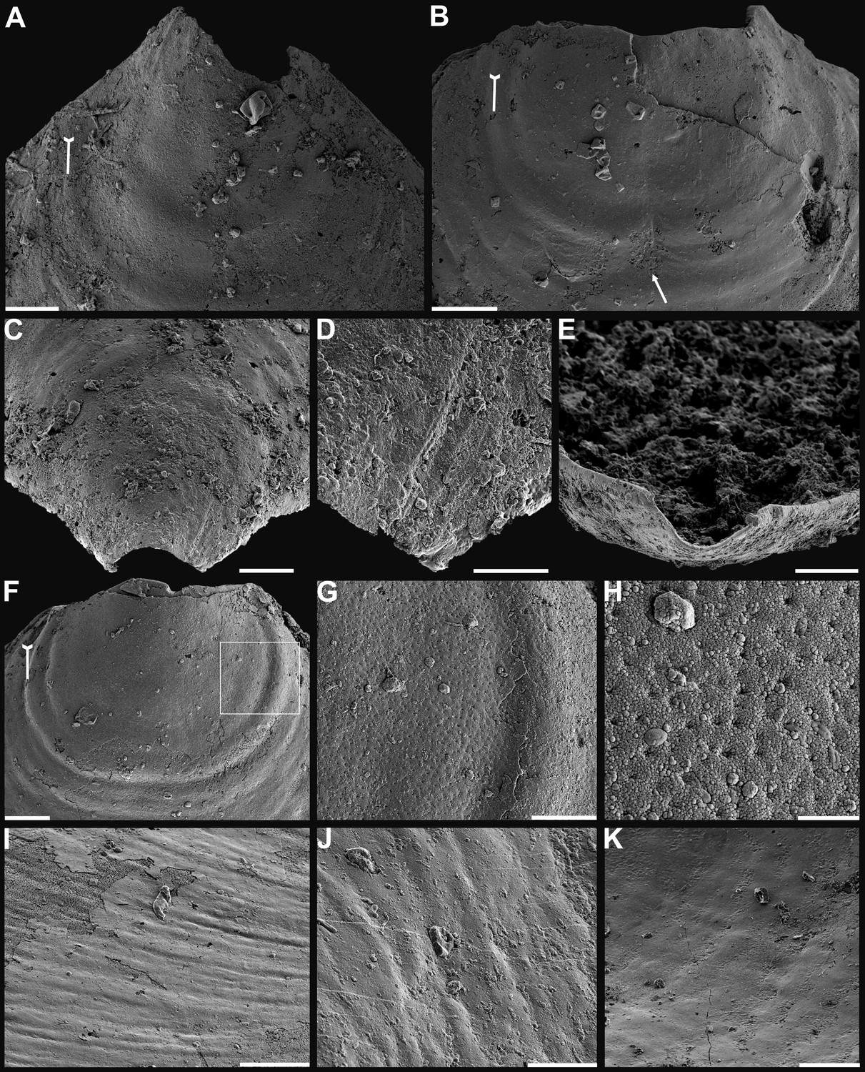
Appendix 1—figure 3
Shell characters and ornamentation of Latusobolus xiaoyangbaensis gen. et sp. nov. from the Cambrian Series 2 Shuijingtuo Formation in southern Shaanxi, South China.
(A) Enlarged ventral metamorphic shell, noting the developed halo by tailed arrow, protegulum by double-headed arrow and brephic lobes by arrows, ELI-XYB S4-3 AU10. (B–C) Ventral valve, ELI-XYB S4-2 BO09. (B) Metamorphic shell of mature valve, noting the halo by tailed arrow, protegulum by double-headed arrow and brephic lobes by arrows. (C) Posterior view, noting the halo by tailed arrow. (D–G) Dorsal valve, ELI-XYB S4-2 BO11. (D) Metamorphic shell, noting the halo by tailed arrow. (E) Lateral dorsal view, noting the halo by tailed arrow, protegulum by double-headed arrow and brephic lobes by arrows. (F) Post-metamorphic pustules, note sparsely packed concentric growth lines by arrows. (G) Enlarged pustules. (H) Enlargement of metamorphic pitted ornament, ELI-XYB S4-2 BO08. Scale bars: (A), (B), (G), 50 µm; (C–E), 100 µm; (F), 200 µm; (H), 5 µm.
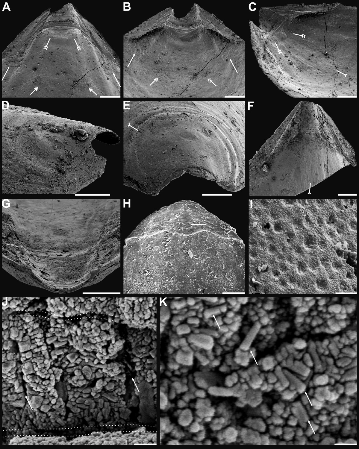
Appendix 1—figure 4
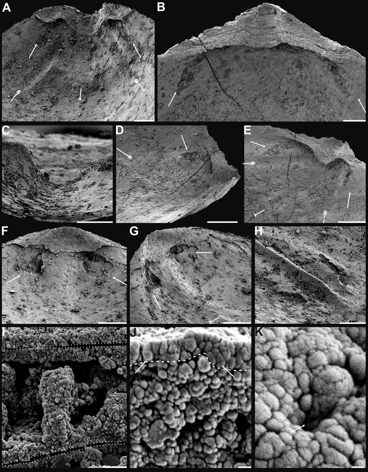
Internal characters and shell ultrastructures of Latusobolus xiaoyangbaensis gen. et sp. nov. from the Cambrian Series 2 Shuijingtuo Formation in southern Shaanxi, South China.
(A–B) Ventral valve, ELI-XYB S4-3 AU10. (A) Interior showing posterolateral muscle scars below propareas by arrows, pedicle nerve by tailed arrow, vascula lateralia by double-headed arrows. (B) Posterior view of pedicle groove. (C–E) Ventral valve, ELI-XYB S5-1 BR09. (C) Interior view showing triangular pseudointerarea and paired posterolateral muscle scars by arrows. (D–E) Lateral view, note elevated pseudointerarea with posterolateral muscles underneath by arrows, pedicle nerve by tailed arrow, vascula lateralia by double-headed arrows. (F–G) Dorsal valve, noting paired umbonal muscle scars by arrows, median ridge by tailed arrow, ELI-XYB S4-2 BO11. (H) Enlargement of terminal of median ridge, ELI-XYB S5-1 BS07. (I) Ventral, enlarged columnar architecture, outlining organic membranes between the stratiform lamellae of stacked sandwich columnar units, ELI-XYB S5-1 BS01. (J) Nanoscale apatite spherules of granule aggregations (arrows) of ventral shell, note primary-secondary layer boundary by dashed line, ELI-XYB S4-2 BO12. (K) Enlarged nanoscale spherules of pitted ornament (arrow) on primary layer, ELI-XYB S4-2 BO08. Scale bars: (A), (B), (H), 50 µm; (C), 100 µm; (D–G), 200 µm; (I), 2 µm; (J), 500 nm; (K), 200 nm.
Appendix 1—figure 5
Ventral and dorsal valves of Eoobolus acutulus sp. nov. from the Cambrian Series 2 Shuijingtuo Formation in Three Gorges areas, South China.
(A–D) Juvenile with weakly developed pseudointerarea, box indicates area in Appendix 1—figure 6D, ELI-WJP 7 CE05. (E–H) Juvenile, note slightly developed pseudointerarea, the halo by tailed arrow, paired posterolateral muscle scars by arrows, pedicle nerve by double-headed arrow, ELI-AJH S05 BT11. (I–K) Mature valve, developed paired posterolateral muscle scars (arrows) underneath elevated pseudointerarea, ELI-AJH S05 BT14. (L–M), Dorsal valve with rudiment of median ridge, ELI-AJH 1-5-01, ELI-AJH 1-5-07. Scale bars: (A–H), (L), (M), 200 µm; (I–K), 400 µm.
Appendix 1—figure 6
Shell characters and ornamentation of Eoobolus acutulus sp. nov. from the Cambrian Series 2 Shuijingtuo Formation in Three Gorges areas, South China.
(A) Enlarged ventral metamorphic shell, noting the halo by tailed arrow, ELI-AJH S05 BT11. (B) Enlarged ventral metamorphic shell, noting the halo by tailed arrow and drape structures outside the halo by arrow, ELI-AJH S05 BT05. (C–E) Ventral valve, ELI-WJP 7 CE05. (C) Posterior ventral view of metamorphic shell. (D) Fine ridges on the margin of protegulum. (E) Posterior view of pedicle groove. (F–H) Ventral valve, ELI-AJH ST 8-2-3 BT04. (F) Metamorphic shell, noting the halo by tailed arrow, box indicates area in G. (G) Metamorphic pits. (H) Enlarged pits. (I) Dense concentric growth lines on ventral external surface, ELI-AJH 8-2-3 BT04. (J) Elongate pustular ornamentation on external surface, ELI-AJH 8-2-3 BT03. (K) Elongate pustule ornamentation on interior, ELI-AJH 8-2-3 BT04. Scale bars: (A–C), (E), (F), (I), (K), 50 µm; (D), (G), 20 µm; (H), 5 µm; (J), 10 µm.
Appendix 1—figure 7
Shell characters and ultrastructures of Eoobolus acutulus sp. nov. from the Cambrian Series 2 Shuijingtuo Formation in Three Gorges areas, South China.
(A–D) Ventral valve, ELI-AJH SJT S05 BT11. (A–C) Interior noting elevated pseudointerarea with posterolateral muscles underneath by arrows, pedicle nerve by tailed arrow, anterior muscle scars by double-headed arrows, umbonal muscle scars by double-tailed arrows. (D) Lateral view of metamorphic shell. (E) Posterior view of ventral metamorphic shell, note the halo by tailed arrow, ELI-AJH SJT S05 BT04. (F–G) Ventral valve, ELI-AJH SJT 8-2-3 BT02. (F) Pseudointerarea of a large valve, noting pedicle nerve by tailed arrow. (G) Posterior view of pedicle groove. (H) Dorsal pseudointerarea, ELI-AJH 1-5-01. (I) Enlarged pitted ornament on primary layer, ELI-AJH 8-2-2 Lin005. (J–K) Ventral valve, ELI-AJH S05 BT12. (J) Apatite spherules of granule aggregations, note organic canals of columns by arrows, outlining organic membranes between the stratiform lamellae of stacked sandwich column units. (K) Enlarged nanoscale spherules of granule aggregations, note elongate rods by arrows. Scale bars: (A–C), (G), (H), 100 µm; (D), 50 µm; (E), (F), 200 µm; (I), 2 µm; (J), 1 µm; (K), 400 nm.
Additional files
-
Supplementary file 1
Average dimensions and ratios of ventral and dorsal valves of Latusobolus xiaoyangbaensis gen. et sp. nov. from the Cambrian Series 2 Shuijingtuo Formation, South China.
- https://cdn.elifesciences.org/articles/88855/elife-88855-supp1-v1.docx
-
Supplementary file 2
Average dimensions and ratios of ventral and dorsal valves of Eoobolus acutulus sp. nov. from the Cambrian Series 2 Shuijingtuo Formation, South China.
- https://cdn.elifesciences.org/articles/88855/elife-88855-supp2-v1.docx
-
MDAR checklist
- https://cdn.elifesciences.org/articles/88855/elife-88855-mdarchecklist1-v1.pdf
Download links
A two-part list of links to download the article, or parts of the article, in various formats.
Downloads (link to download the article as PDF)
Open citations (links to open the citations from this article in various online reference manager services)
Cite this article (links to download the citations from this article in formats compatible with various reference manager tools)
Evolution and diversity of biomineralized columnar architecture in early Cambrian phosphatic-shelled brachiopods
eLife 12:RP88855.
https://doi.org/10.7554/eLife.88855.4


